 A human embryo model replicates key early developmental processes and generates organ-seed cells in vitro. [Photo provided to chinadaily.com.cn]
Chinese researchers have developed a first-of-its-kind human embryo-disc model capable of providing a controllable platform for generating organ-seed cells in vitro, laying the groundwork for future organ cultivation and regenerative medicine therapies. The breakthrough was published in the journal Cell on Wednesday.
International guidelines prohibit culturing intact human embryos beyond 14 days after fertilization, a stage when gastrulation — the process that gives rise to all organ precursors — begins and marks the start of organ formation, one of the most critical events in human development.
This makes laboratory-created embryo-like models the only ethical way to study the decisive 14-to-21-day window of early human development and explore what is often described as the "black box" of embryology.
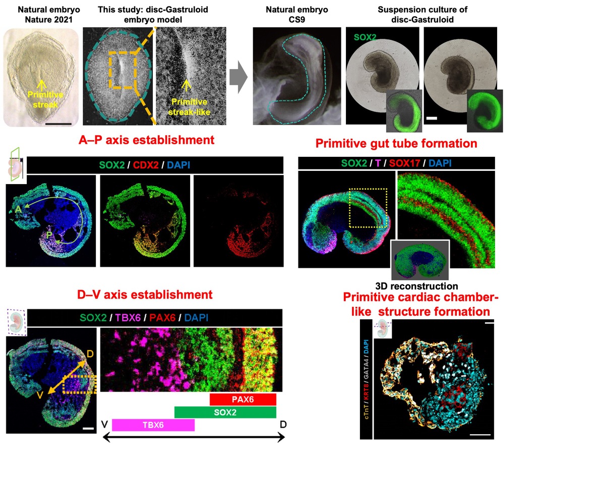
"Gastrulation is when the body's basic architecture is established, transforming the embryo from a flat disc into a three-dimensional structure," said Yu Leqian, corresponding author of the study and a professor at the Institute of Zoology of the Chinese Academy of Sciences.
While the stage is crucial, Yu noted that previous human embryo models could generate only certain cell types but failed to form the primitive streak — a narrow groove that serves as the gateway to this transformation. As a result, cellular organization remained random and uncontrollable, hindering further mechanistic investigations.
To address this challenge, the research team adopted a bioengineering approach to constructing human embryo models. Drawing on high-resolution maps from their previous work that detailed the spatial positions and signaling environments of early human cells, researchers developed a microfabricated culture device that positioned different cell types at precisely defined locations.
The approach replicated the spatial architecture of natural embryos, ensuring that organ-seed cells emerged in the correct positions.
The resulting models, named "disc-Gastruloids", initiated gastrulation and formed primitive-streak-like structures — a feat beyond the reach of previous laboratory models. This triggered large-scale, coordinated cell migration across the surface of the disc, reproducing the hallmark dynamics of early development.
More than 80 percent of the models successfully replicated key developmental processes. Over a seven-day culture period, they formed a neural tube, a primitive gut containing lung, liver and pancreas progenitors, and a primitive heart chamber exhibiting autonomous rhythmic contractions. Single-cell analysis confirmed that their cellular composition closely matched that of a natural 21-day-old human embryo.
"The study has laid the foundation for the ultimate goal of large-scale, modular production of organ-seed cells in vitro to support organ manufacturing and regenerative medicine — potentially enabling tissue repair or even the construction of organs in the laboratory," Yu said.
He emphasized that if such organs are eventually produced, they would face far fewer ethical constraints because the human embryo models are essentially a complex form of organoid composed of functional cells rather than a living organism. The same applies to organs generated from the organ-seed cells.
Data from the China National Organ Donation and Transplantation Committee showed that an estimated 300,000 patients in China require organ transplants each year, but fewer than 20,000 procedures are performed. The shortage is not unique to China. Globally, only one in 10 patients in need receives a transplant.

